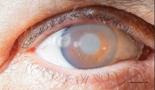
What is Glaucoma ?

Glaucoma


Glaucoma is not a single disease process but a group of disorders characterized by a progressive optic neuropathy resulting in a characterstic appearance of the optic disc and a specific pattern of irreversible visual field defects that are associated frequently but not invariably with raised intraocular pressure (IOP).


 Thus, IOP is the most common risk factor but not the only risk factor for development of glaucoma.


Consequently the term ‘ocular hypertension’ is used for cases having constantly raised IOP without any associated glaucomatous damage. Conversely, the term normal or low tension glaucoma (NTG/LTG) is suggested for the typical cupping of the disc and/or visual field defects associated with a normal or low IOP.


Increase intraocular pressure , increase disc diameter, and decrease peripheral vision is called as glaucoma


Classification


Clinico-etiologically glaucoma may be classified as follows:


(A) Congenital and developmental glaucomas


1. Primary congenital glaucoma (without associated anomalies).


2. Developmental glaucoma (with associated anomalies).


(B) Primary adult glaucomas


1. Primary open angle glaucomas (POAG)

2. Primary angle closure glaucoma (PACG)

3. Primary mixed mechanism glaucoma


(C) Secondary glaucomas


PATHOGENESIS OF GLAUCOMATOUS OCULAR DAMAGE


As mentioned in definition, all glaucomas (classified above and described later) are characterized by a progressive optic neuropathy.


 It has now been recognized that progressive optic neuropathy results from the death of retinal ganglion cells (RGCs) in a typical pattern which results in characteristic optic disc appearance and specific visual field defects.


Pathogenesis of retinal ganglion cell death


Retinal ganglion cell (RGC) death is initiated when some pathologic event blocks the transport of growth factors (neurotrophins) from the brain to the RGCs.


The blockage of these neurotrophins initiate a damaging cascade, and the cell is unable to maintain its normal function. The RGCs losing their ability to maintain normal function undergo apoptosis and also trigger apoptosis of adjacent cells. 


Apoptosis is a genetically controlled cell suicide programme where by irreversibaly damaged cells die, and are subsequently engulfed by neighbouring cells, without eliciting any inflammatory response.


Retinal ganglion cell death is, of course, associated with loss of retinal nerve fibres. As the loss of nerve fibres extends beyond the normal physiological overlap of functional zones. The characteristic optic disc changes and specific visual field defects become apparent over the time.


OPTOMETRY-SHARP VISION

Optometrist

【 About Ophthalmology 】 【 Adie's Pupil 】 【 Adult Strabismus 】 【 Age-Related Macular Degeneration 】 【 Albinism 】 【 Alzheimer’s Disease, Dementia and the Eye 】 【 Amblyopia 】 【 Anisocoria 】 【 Antibiotic Eye Drops 】 【 Anti-VEGF Treatments】 【 Aqueous Humor 】 【 Arcus Senilis 】 【 Artificial Tears 】 【 Astigmatism 】 【 Avastin 】 【 Bacterial Keratitis 】 【 Bell's Palsy 】 【 Black Eye 】 【 Blepharitis 】 【 Blocked Tear Duct 】 【 Blood in Eye 】 【 Blood Pressure 】 【 Bloodshot Eye 】 【 Blue Light 】 【 Blurriness 】 【 Botulinum Toxin (Botox) for Facial Wrinkles 】 【 Bowman's membrane 】 【 Branch Retinal Vein Occlusion (BRVO) 】 【 Burning Eyes 】 【 Cancer of the Eye 】 【 Carotid Arteries 】 【 Carotid Artery Disease 】 【 Cataract and Glaucoma Surgeries 】 【 Combined 】 【 Cataracts 】 【 Cellulitis 】 【 Central Retinal Vein Occlusion 】 【 Central Serous Chorioretinopathy 】 【 Cerebrospinal Fluid (CSF) 】 【 Chalazia and Stye 】 【 Charles Bonnet Syndrome 】 【 Choroid 】 【 Choroidal Neovascular Membranes 】 【 Chronic Angle-Closure Glaucoma 】 【 Ciliary Body 】 【 Coloboma 】 【 Color Blindness 】 【 Color Vision 】 【 Colored Rings in the Iris 】 【 Colors look dull, faded or different 】 【 Computer Use 】 【 Cones 】 【 Conjunctiva 】 【 Conjunctivitis (Pink Eye) 】 【 Contact Lens Care 】 【 Contact Lenses 】 【 Contact Lens-Related Eye Infections Cornea 】 【 Corneal Abrasion 】 【 Corneal Cross-Linking 】 【 Corneal Dystrophies 】 【 Corneal Erosion 】 【 Corneal Inlays 】 【 Corneal Laceration 】 【 Corneal Topography 】 【 Corneal Transplantation 】 【 Corneal Ulcer 】 【 Cosmetic Contact Lenses 】 【 Crossed Eyes (Strabismus)】 【 Crow's Feet 】 【 Crusty Eyelid or Eyelashes 】 【 Cytomegalovirus Retinitis 】 【 Dark Curtain in Vision 】 【 Dark Spots in Vision 】 【 Dementia and the Eye 】 【 Depth Perception 】 【 Descemet's Membrane 】 【 Detached Retina 】 【 Diabetic Eye Disease 】 【 Diabetic Retinopathy 】 【Digital Devices and Your Eyes 】 【 Dilated Pupil 】 【Dilating Eyedrops】 【Discharge From Eye】 【Distorted Vision】 【Double Vision】 【Drainage Angle】 【Drooping Eyelid】 【Drusen】 【Dry Eye】 【Dry Eye Tips】 【Dryness】 【Ectropion】 【Endophthalmitis】 【Endothelium】 【Enlarged Pupil】 【Entropion】 【Enucleation】 【Epithelium】 【Erectile Dysfunction Medication】 【Evisceration】 【Excessive Blinking in Children】 【Eye Allergies】 【Eye Anatomy Overview】 【Eye Cancer】 【Eye Color】 【Eye Donation】 【Eye Drops】 【Eye Exams】 【Eye Injections】 【Eye Injuries】 【Eye Lymphoma】 【Eye Muscles】 【Eye Pressure】 【Eye Removal Surgery】 【Eyelashes】 【Eyelashes and Cosmetics】 【Eyelid】 【Eyelid Droop】 【Eyelid redness and rash】 【Eyelid rough, red and swollen on inside】 【Eyelid Spasm and Twitching】 【Eyelid Surgery】 【Eyelid Turns Out】 【Eyes with different colored irises】 【Eyestrain (Condition)】 【Eyestrain (Symptom)】 【Eylea】 【Face-Down Recovery After Retinal Surgery】 【Farsightedness (hyperopia)】 【Feeling of Something in Eye】 【Flashes of Light】 【Floaters and Flashes】 【Floaters in Vision】 【Fluorescein Angiography】 【Food and Nutrition】 【Fovea】 【Fuchs’ Dystrophy】 【Fundus】 【Fungal Keratitis】 【Giant Cell Arteritis】 【Giant Papillary Conjunctivitis】 【Glasses】 【Glaucoma】 【Glaucoma and Cataract Surgeries】 【Combined】 【Gonioscopy】 【Graves' Disease】 【Grittiness】 【Halos Around Lights】 【Headache and Eye Problems】 【Headache Behind Eye】 【Headache: Eye Conditions That Can Cause】 【Headache】 【Hemangioma】 【Herpes Keratitis】 【Herpes Zoster (Shingles)】 【Heterochromia】 【Histoplasmosis】 【HIV/AIDS and the Eye】 【Hyperopia (farsightedness)】 【Hyphema】 【Idiopathic Intracranial Hypertension】 【Inflammation, General】 【Interferon】 【Intraocular Implants (IOLs)】 【Intraocular Pressure】 【Iridocorneal Endothelial Syndrome】 【Iris】 【Irritation】 【Ischemic Optic Neuropathy】 【Itchiness】 【Juvenile Idiopathic Arthritis Uveitis】 【Juvenile Macular Dystrophy】 【Keratoconus】 【Lacrimal Gland】 【LASIK】 【Latisse (Bimatoprost Ophthalmic Solution)】 【Lattice Degeneration】 【Lazy Eye (Amblyopia)】 【Lens】 【Lens Capsule】 【Light Sensitivity】 【Limited Movement of Eyelid】 【Limited Movement of Eyes】 【Low Vision】 【Lubricating Eye Drops】 【Lucentis】 【Lump on Eyelid】 【Macula】 【Macular Edema】 【Macular Hole】 【Macular Pucker】 【Macular Telangiectasia】 【Marfan Syndrome】 【Marijuana】 【Meibomian Glands】 【Microbiome of the Eye】 【Microvascular Cranial Nerve Palsy】 【Migraine】 【Mohs Surgery】 【Monovision (Blended Vision)】 【Myasthenia Gravis】 【Myopia】 【Narrow Angles】 【Nearsightedness (Myopia)】 【Neuropathic Corneal Pain】 【Nevus】 【Night Vision】 【Night Vision Problem】 【Nystagmus】 【Objects Appear Smaller or Different Sizes】 【Ocular Hypertension】 【Ocular Melanoma】 【Ocular Rosacea】 【Onchocerciasis (African River Blindness)】 【Optic Disc】 【Optic Nerve】 【Optic Neuritis】 【Optical Coherence Tomography】 【Orbital Fracture】 【Pain Around Eye】 【Pain Behind Eye】 【Pain in Eye】 【Parts of the Eye】 【Peripheral Retina】 【Photokeratitis】 【Photoreceptors】 【Pigment Dispersion Syndrome】 【Pinguecula and Pterygium】 【Pink Eye (Conjunctivitis)】 【Pink Eye (symptom)】 【Plaquenil】 【Posterior Capsulotomy】 【Posterior Vitreous Detachment】 【Pregnancy】 【Presbyopia】 【Pseudoexfoliation Syndrome】 【Pseudostrabismus】 【Ptosis】 【Pupil】 【Red Eye】 【Red or purple raised legion on or near the】 【eye or eyelid 【Redness-Relieving Eye Drops】 【Reduced Vision】 【Refractive Surgery】 【Retina】 【Retinal Artery Occlusion】 【Retinal Detachment】 【Retinitis Pigmentosa】 【Retinoblastoma】 【Retinopathy of Prematurity】 【Ring Around the Cornea】 【River Blindness (Onchocerciasis)】 【Rods】 【Scarring in or on the eyeball】 【Sclera】 【Scleritis】 【Scratched Eye】 【Screen Use and Safety】 【Secondary Cataract】 【Shadow or Dark Curtain in Vision】 【Shingles (Herpes Zoster)】 【Sjögren's Syndrome】 【Sleep Crust】 【Slit Lamp】 【Small Incision Lenticule Extraction】 【(SMILE)】 【Small pupils】 【Solar Eclipses】 【Spasm/Twitching】 【Sports Eye Safety】 【Spot on Eye, Brown】 【Spot on Eye, Cloudy or White】 【Spot on Eye, Red or Pink】 【Spot on Eye, Yellow】 【Spot on Eyelid, Colored】 【Squinting】 【Starbursts Around Lights】 【Stargardt Disease】 【Steroid Tablets】 【Stickler Syndrome】 【Strabismus (Crossed Eyes)】 【Straight Lines Look Bent or Wavy】 【Stroma】 【Stye and Chalazia】 【Subconjunctival Hemorrhage】 【Sun and UV Light】 【Sunglasses】 【Swelling Around Eye】 【Swollen Eye】 【Tear Duct】 【Tear Film】 【Tearing】 【Tears and Crying】 【Torn Retina】 【Toxoplasmosis】 【Trachoma】 【Transplantation and the Eye】 【Travel and Eye Health】 【Trichiasis】 【Trichotillomania】 【Tunnel Vision】 【Two Different Colored Eyes/Irises】 【Usher Syndrome】 【Uvea】 【Uveitis】 【Veterans】 【Vision Loss, Central】 【Vision Loss, General】 【Vision Loss, Peripheral (Side)】 【Vision loss, temporary】 【Vitamin A Deficiency】 【Vitrectomy Surgery】 【Vitreomacular Traction】 【Vitreous】 【When Can I Resume Exercise?】 【Zonules】
Previous Post Next Post
//disable Text Selection and Copying