Visual Field
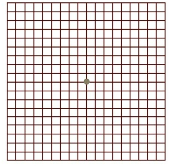
This test should be done to almost all patients. An Amsler chart is used for central scotoma patients and the Goldman’s perimeter for peripheral field of vision.
A Goldman perimeter is the best way to estimate the field defect and classify the peripheral defects. The perimetry report
indicates the extent of the scotoma, their position and density.
Peripheral field defects are found in commonly in glaucoma, retinitis pigmentosa patients.
Automated perimeters though more accurate, seldom reveal the true threshold values, due to poor fixation and visual acuity.
The central scotoma will explain the ability of the patient as to why he is able or unable to read the text.
Central scotoma the larger they are, the more the magnification is needed and difficult to achieve 1 M visual acuity.
If the scotoma is to the side of the fixation target it becomes easier to achieve the visual acuity.
The Amsler grid should always be at hand and presented to all patients on routine.
The recording of the scotoma also forms a base or guide for the follow-up visits.
Some central scotoma patients will fix eccentrically, this is a healthier sign as the eccentric fixation developed indicates better
prognosis.
Central field loss often indicates difficulty with visual skills for reading, whereas peripheral field loss often indicates difficulty in mobility.
(The position and the extent of the scotoma affect the prognosis and manifestations of patient’s complaints- e.g. a dense scotoma to the right of fixation may cause portions of words to disappear. A scotoma below may lead to problem of finding the beginning of the line of the text. A very dense large scotoma may show no improvement with devices. )
Tags:
OPHTHALMIC PROCEDURES

