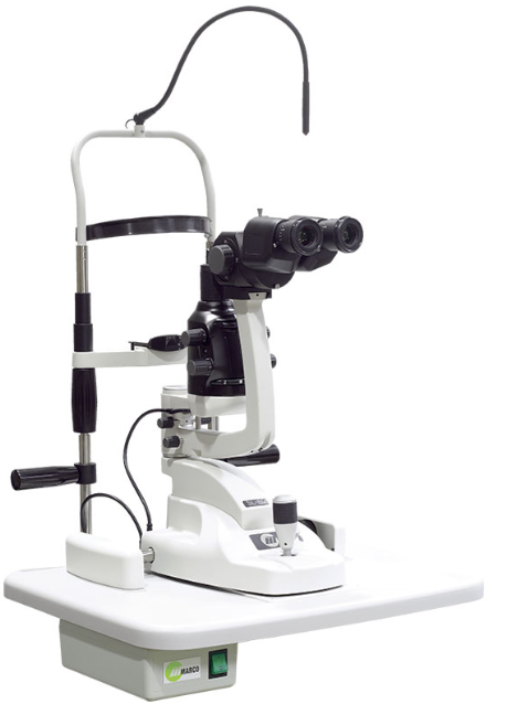
Slit lamp
Slit lamp is most commonly used by optometrist, ophthalmologist, Eye doctor and eye care service provider. It is also called as slit lamp biomicroscope because in this device we can see with both eye at time. This instrument is very easy to used, handy and very essential in ophthalmic practice during eye examination. It is one of the basic instrument used for eye examination, diagnosis of disease.
Slit lamp test basically do for examination and diagnosis of disease with full Hd view of eye. It is also used in detect the glaucoma and it's type. The angle of anterior chamber either closure or open its measure with help of slit lamp.
Slit lamp can not damage the eye because it's not directly contact to your eye. In some case like long time eye examination process lead to photophobia, dryness, lacrimation, irritation, Diminished vision for short time, light hallows this symptoms may be happened for very shot period.
Slit lamp also used for examine the retina and retinal disease with the help of 90D lens lens. The ophthalmologist and optometrist can see retina through 90 D lens. It is also called as funduscopy. It is not used for burned retina heal. There is another equipment and instrument used for burn retina heal is called as argon laser machine.
Eye part
During slit lamp test, the eye doctor will examine all part of your eye, including the:
- Eyelids
- Conjunctiva
- Iris
- Lens
- Sclera
- Cornea
- Retina
- Optic nerve
- Choroid
- Macula
Eye diseases
Slit lamp test can diagnose the different eye disease including:
1. Eyelid diseases:
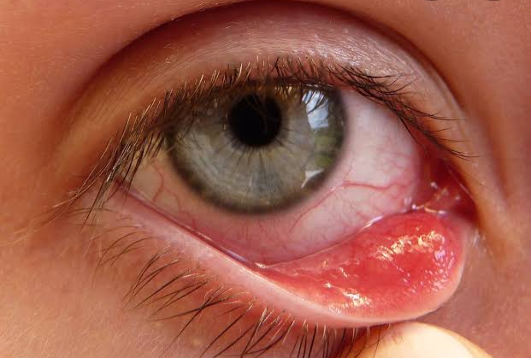
- Chalazion
- Style
- Internal hordeolum
- External hordeolum
- Ptosis
- Blepharitis
- Symplempharon
- Ankyloblempharon
- Poliosis
- Madrosis
2. Corneal diseases:
- Corneal foreign body (FB)
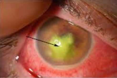
- Corneal ulcer
- Bacterial corneal ulcer
- Viral corneal ulcer
- Fungal corneal ulcer
- Allergic corneal ulcer
- Purulent corneal ulcer
- Mucopurulent corneal ulcer
- Microcornea
- Megalocornea
- Keratoconus
- Keratoglobus
- Corneal opacity
3. Scleral diseases:
- Scleritis
- Episcleritis
- Blue sclera
- Traumatic injury
4. Conjunctival diseases:
- Conjunctivitis
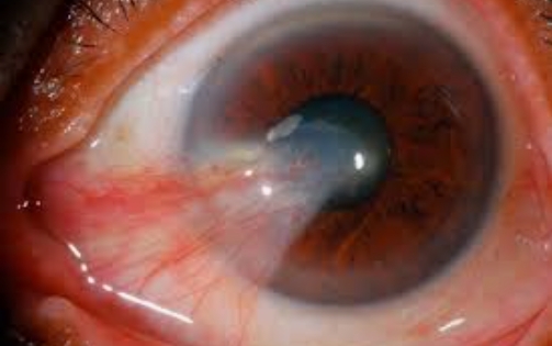
- Pterygium
- Traumatic injury
5. Lens diseases:
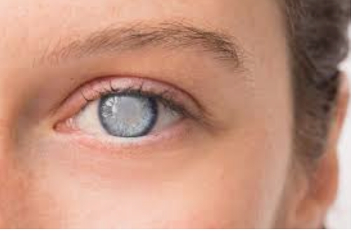
- Cataract
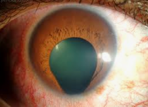
- Dislocation of lens
- Subluxation of lens
- Weak zonules
- Lens coloboma
6. Iris diseases:
- Iris coloboma
- Irities
- Iridodonesis
- Iridocyclities
7. Retinal diseases:
- Retinities
- Macular oedema
- CRVO
- CRAO
- BRAO
- BRVO
- Diabetic retinopathy
- Hypertensive retinopathy
- Retinal detachment
Techniques of examination
Techniques of slit lamp examinations for various parts of eye. There are 7 basic method of slit lamp examinations.
- Diffuse illumination
- Direct illumination
- Indirect illumination
- Retro illumination
- Oscillatory illumination
- Specular reflection
- Sclerotic scatter.
OPTOMETRY-SHARP VISION
Optometrist

