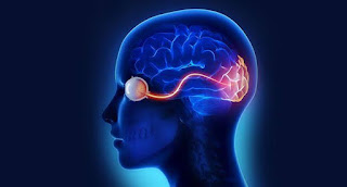
Important term & Points in neurophthalmology
Suprageniculate lesions of visual pathway usually produce visual field defects with macular sparing.
• Optic nerve lesions produce negative scotomas whereas macular lesions cause positive scotoma.
• Chromophobe adenoma is the most common primary intracranial tumour producing neuro-ophthalmological features.
• Gaze evoked amaurosis is seen in optic nerve sheath meningioma.
• Horner’s syndrome (lack of sympathetic innervation) is characterized by miosis, mild ptosis, mild enophthalmos, anhydrosis of the face on the affected side.
Loss of cilio-spinal reflex and heterochromia (ipsilateral iris is of light colour).
Tests to confirm diagnosis of Horner’s syndromeare: dilation lag, and cocaine test (normal pupil dilates while Horner’s pupil does not dilate with topical cocaine).
• The swelling of the optic disc in papillitis rarely exceeds 2-3D.
• Scintilating scotoma is a feature of migraine.
• Unilateral central scotoma is the earliest symptom of compression of optic nerve.
• Hippus (alternate rhythmatic dilation and constriction of pupils) is a feature of multiple sclerosis.
• Erythropsia (red coloured vision) may be experienced by some patients after cataract extraction.
• Pupil sparing, third nerve paralysis suggests a medical cause (diabetes or hypertension). While in surgical causes (aneurysm, tumour) pupil is also invovled.
• The two most common ocular signs of myasthenia gravis are ptosis and extraocular muscle weakness (paralytic squint).
• Neuromyelitis optica (Devic’s disease) may be associated with sudden bilateral blindness.
• Papillitis and retrobulbar neuritis :
Painful ocular movement is more common in retrobulbar neuritis than papillitis and fundus is normal in retrobulbar neuritis while papillitis has characteristic fundus abnormalities.

